Fig. 2.
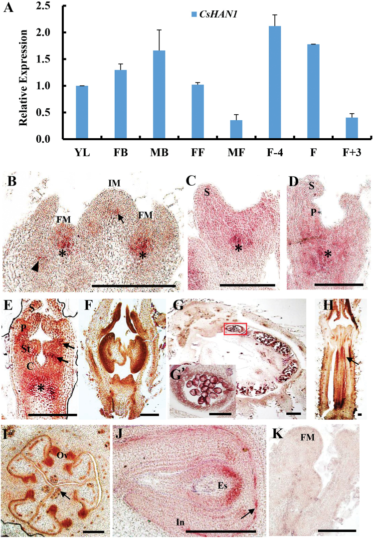
Expression analysis of CsHAN1 in cucumber. (A) Quantitative RT–PCR (qRT–PCR) analysis of CsHAN1 in different tissues of cucumber. YL, young leaves; FB, female buds; MB, male buds; FF, female flowers; MF, male flowers; F-4, young fruits 4 d before anthesis; F, fruit at anthesis; F+3, fruits 3 d after anthesis. The Ubiquitin extension protein (UBI-ep) gene was used as an internal reference to normalize the expression data. (B–K) In situ hybridization with the CsHAN1 antisense probe (B–J) and sense probe (K). (B) In the ucumber shoot apex, CsHAN1 is expressed in the junction region of the inflorescence meristem (IM) and stem (arrow), the junction regions of the floral meristem (FM) and stem (asterisk), and the axil of leaf primordia (arrowhead). (C–E) Floral buds at stage 2 (C), 3 (D), and 4 (E). Asterisks show the expression domain of CsHAN1 at the junction of the meristem and stem, and arrows indicate the expression of CsHAN1 at the boundary between the petal and stamen, and the boundary between the stamen and initiating carpel primordia. (F–G’) Male flowers at stage 9 (F) and stage 11 (G); (G’) is a high magnification view of the anther in (G). The signal of CsHAN1 was detected in the developing anther, tapetum cell layer, and the uninuclear pollen. (H) Female flower in stage 8. CsHAN1 is expressed in the ovary (arrow). (I, J) Cross-sections of the female ovary in stage 9 (I) and stage 10 (J) showing the expression domain of CsHAN1 in the ovules and the base of the embryo sac. (K) No signal was found on hybridization with the sense CsHAN1 probe. S, sepal; P, petal; St, stamen; C, carpel; Ov, ovule; In, integument; Es, embryo sac. Bar=100 μm. (This figure is available in colour at JXB online.)

