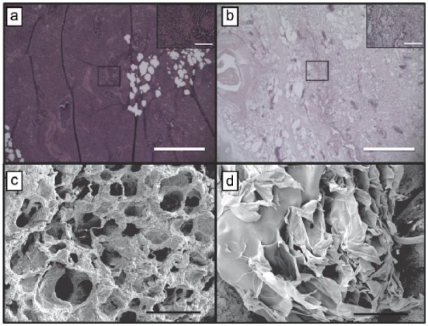
Figure 3.

Characterization of decellularization. Hematoxylin and eosin staining at (a) day 0 and (b) day 7 confirm complete decellularization following SDS baths (scale bar, 150 µm; inset 40× magnification; scale bar, 20 µm). (c) Well-formed and cellularized tubular vascular structures are in lung tissue prior to processing (scale bar, 100 µm). (d) Decellularized vascular structures retain tubular morphology and architecture (scale bar, 100 µm).
