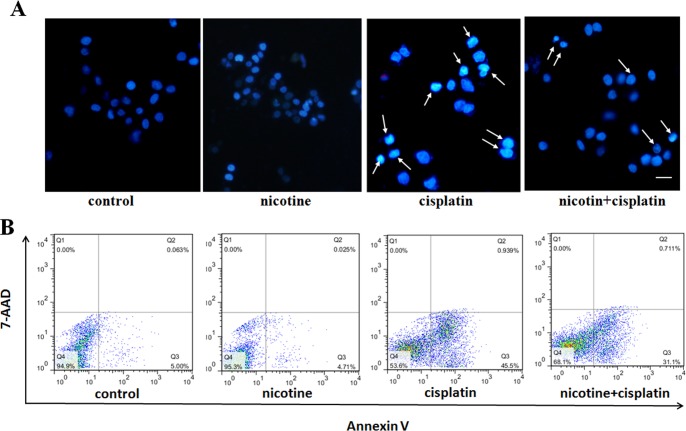
Fig 3. Nicotine inhibition of cisplatin-induced apoptosis.
A: Cisplatin induced cells apoptosis compared with the control. In contrast, the cells co-treated with 100 μM nicotine and 20 mM cisplatin showed little nuclear fragmentation and few apoptotic bodies by fluorescence microscopy (200 X); B: Cisplatin induced apoptosis was assayed by AnnexinV/7-AAD staining. Flow cytometry plots showed that the proportion of total apoptotic cells was decreased when exposed to 20 mM cisplatin combined with 100 μM nicotine.

