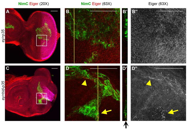
Figure 6. Hemocytes are a source of the TNF ligand Eiger.
Shown are eye-antennal imaginal discs from third instar larvae labeled with anti-Eiger antibody (red) and NimC antibody (green) to identify hemocytes (plasmatocytes). Scale bars 50 μm.
(A,B) Anti-Eiger labeling (red) of control (ey>p35) eye discs with attached hemocytes (green). Inset from (A) is magnified in (B). Yellow line in (B) marks the orthogonal (YZ) section shown in (B′). Diffuse Eiger staining is seen in disc epithelium, but not in hemocytes (B″).
(C,D) Anti-Eiger labeling of undead (ey>hid-p35) eye discs with attached hemocytes (green). Inset from (C) is magnified in (D). Yellow line in (D) marks the orthogonal (YZ) section shown in (D′). At higher magnification, increased Eiger labeling can be seen in hemocytes in the overgrown region of an undead disc (arrows in D,D′,D″) as well as at epithelial cells close to hemocytes (arrowhead in D and D″).

