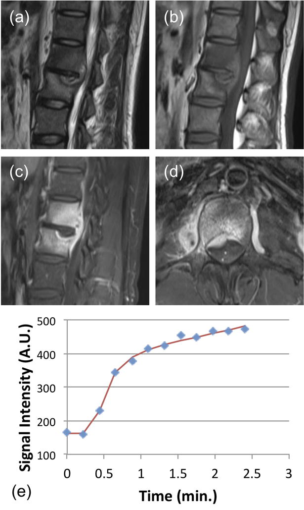
Figure 3.

A 38-year-old male patient with confirmed pathological diagnosis as tuberculosis. (a) MR T2WI and (b) MR T1WI show steolytic destruction in L1–2 vertebral body. A soft tissue mass compressing the spinal canal is shown. The narrowing of intervertebral space is noted, but the signal is not increased. (c) and (d) Contrast-enhanced MR T1WI shows a heterogeneously enhanced lesion. (e) The DCE kinetics shows a clear persistent enhancement pattern. The signal at the last time point compared to the 1-min time point shows 20% increase. In pharmacokinetic analysis, the fitted Ktrans=0.057/min, kep=0.086/min. The persistent enhancement pattern with narrowing of intervertebral space may be used to help diagnosis as TB.
