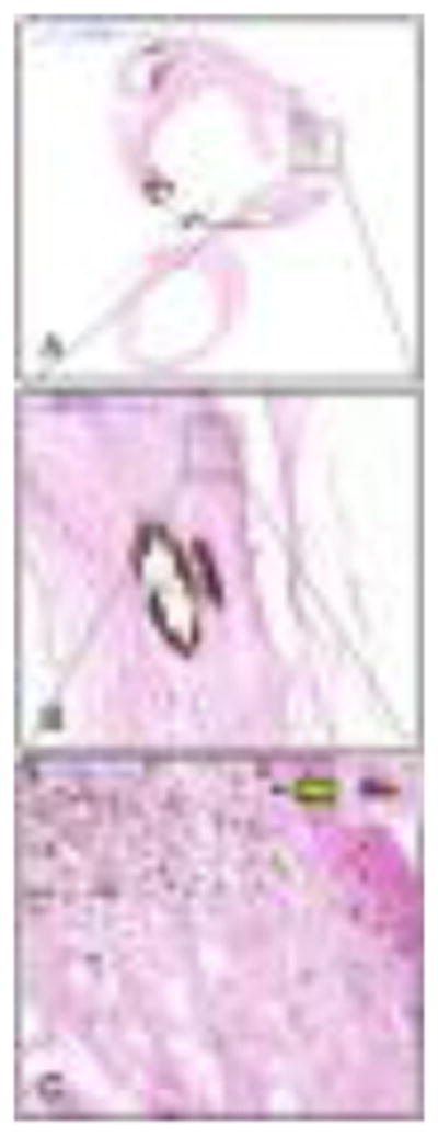
Figure 6.

A representative von Kossa stained CEA tissue section of the bulb segment as show at different magnifications to illustrate the very-small calcified particles (bar, A: 5000μm, B: 500μm and C: 50μm). The very small calcified particles are rounded in shape, but some are coalescing with each other to form a bigger particle (green arrow insert).
