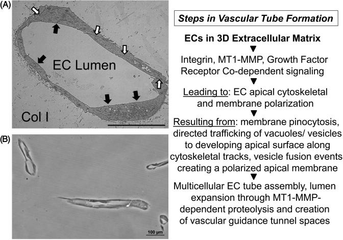
Figure 3. Molecular events controlling EC lumen formation during “Factor”-induced EC tube assembly in 3D collagen matrices.
ECs were seeded as single cells and allowed to form tubes over 120 hr prior to fixation and processing for transmission electron microscopy (A) or plastic thin sectioning (B) to demonstrate EC lumen formation following cross-sectioning of 3D collagen gels. Black arrows indicate the EC apical surface; white arrows indicate EC junctional contacts, Col I indicates the 3D collagen type I matrix. Key regulatory steps that control the EC lumen formation process are highlighted in the right panel. Bar equals 10 μm (A); Bar equals 100 μm (B).

