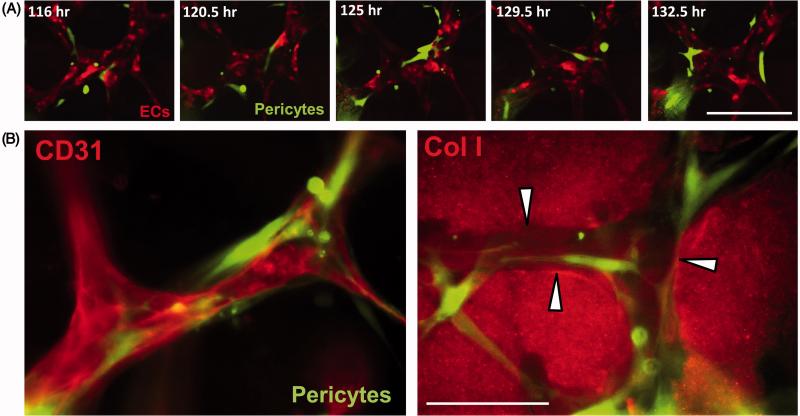
Figure 4. Capillary tube assembly in 3D collagen matrices and role of vascular guidance tunnels in EC-pericyte tube co-assembly.
(A) ECs were co-cultured with GFP-pericytes and at the indicated times, cultures were fixed with paraformaldehyde and stained using anti-collagen type I (Col I) antibodies, and were photographed using light or fluorescent microscopy. EC tubes form, vascular guidance tunnels appear and pericytes are observed to recruit to these tubes on the EC tube abluminal surface within vascular guidance tunnels. Arrowheads indicate the borders of vascular guidance tunnels. Bar equals 50 μm. (B) ECs were co-cultured with GFP-pericytes and at the indicated times, cultures were fixed, immunostained with anti-collagen type I antibodies and photographed using fluorescence microscopy. Marked recruitment of pericytes is observed to EC-lined tubes which are present within vascular guidance tunnels over time. Arrowheads indicate the borders of vascular guidance tunnels. Bar equals 100 μm.

