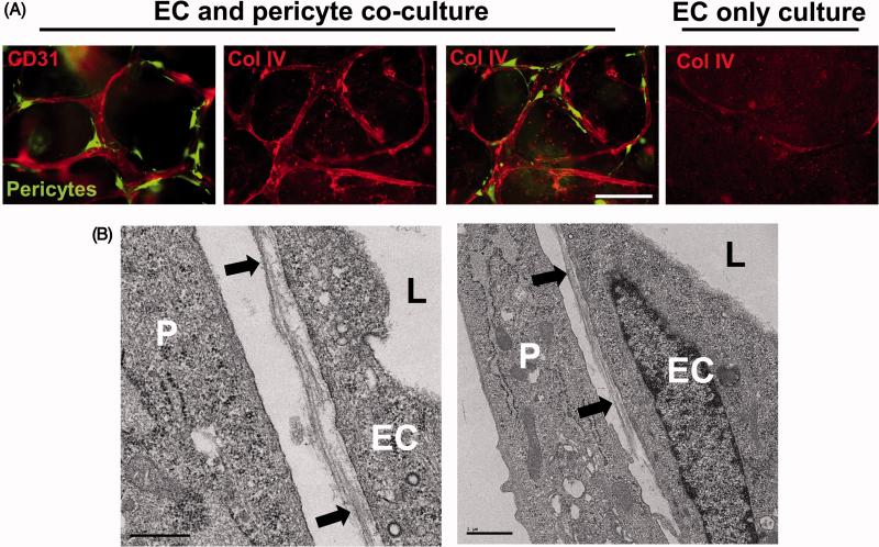
Figure 6. EC-pericyte tube co-assembly leads to vascular basement membrane matrix deposition.
(A) ECs alone or ECs and GFP-pericytes were cultured for 120 hr and then fixed and stained for CD31 or collagen type IV (Col IV), a key basement membrane component. Fixed cultures were not permeabilized with detergent, so that only collagen type IV that was deposited extracellularly is observed. Note that marked basement membrane deposition, as indicated by collagen type IV deposition, is observed only when ECs and pericytes are cultured together. Bar equals 50 μm. (B) EC-pericyte co-cultures were examined by transmission electron microscopy and vascular basement membrane deposition is observed between the two cell types (black arrows). P indicates pericytes, EC indicates endothelial cells, and L indicates the luminal space. Left image- Bar equals 0.5 μm; Right image- Bar equals 1 μm.

