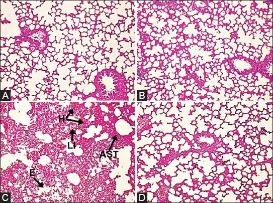
FIGURE 2.

Microphotographs of lung tissues in the study groups. (A,B) Hematoxylin and eosin staining (H&E x 100) revealed that there was no any change in the control and Ukrain groups, (C) Severe edema, hemorrhage, leukocyte infiltration, and alveolar septal thickening were observed in the I/R group. The “H” denotes hemorrhage, “LI” denotes leukocyte infiltration, “AST” denotes alveolar septal thickening, and “E” denotes edema. (D) The microscopic views of lung tissues were near normal in the I/R with Ukrain group. Minimal hemorrhage, leukocyte infiltration, alveolar septal thickening, and a comparatively preserved pulmoner architectures were observed in the I/R with Ukrain group. The slides were graded according to a scoring system previously described by Yamanel et al. [17].
