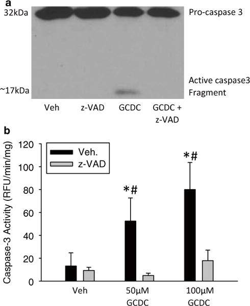
Fig. 2.

Bile acid-induced apoptosis in rat hepatocytes. Hepatocytes were isolated from rats and exposed to vehicle (i.e. 0.1 % dimethyl sulfoxide), 50 μM, or 100 μM GCDC for 6 h with and without a 30 min pretreatment with 10 μM of the pan-caspase inhibitor z-VAD-FMK. Caspase-3 activation after 50 μM GCDC was measured by immunoblot analysis (a) and through a fluorogenic caspase activity assay (b) (*p < 0.05 vs. control; #p < 0.05 vs. matched z-VAD-fmk treated sample)
