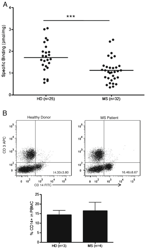
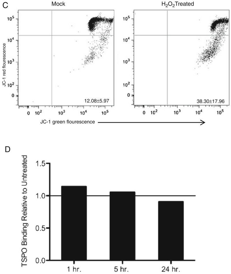
Fig. 2.
MS patients show significantly lower peripheral TSPO levels than HD, which is not due to acute mitochondrial injury. a Using a [3H]PBR28 radioligand binding assay, TSPO was measured in both HD (n=25) and MS patients (n=32), and Bmax is plotted as the pmol of specific binding normalized to protein concentration. The HD mean Bmax is found to be 1.7±0.69 pmol/mg, while the MS patient mean Bmax is found to be 1.1±0.55 pmol/mg. There is significantly lower TSPO expression in the MS cohort, students t-test p=0.0007. b Total PBMC from 3 HD and 4 MS patients were stained with CD14-FITC and CD3-APC antibodies and analyzed by flow cytometry. Representative plots of MS patient and HD cell profiles are shown along with a graph showing the mean ± SD percentage of CD14+ cells in PBMC for each cohort (HD= 14.33±3.80 %, MS=16.46±8.67 %), there are no significant differences seen in the percentages of each cell population. C. Representative plot, n=3, of CD14+ cells untreated and treated with H2O2 for 1 h. The lower right quadrant indicates mitochondrial membrane potential dysfunction, which was 12.08±5.97 % for mock, and 38.30±17.96 % for H2O2 treated. D. A radioligandbinding assay was used to measure specific binding of [3H]PBR28 to H2O2 treated PBMC relative to untreated. The relative amounts are 1.14 after 1 h, 1.06 after 5 h, and 0.91 after 24 h of treatment


