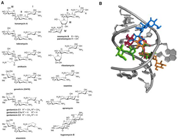
Figure 1.
A) Structures of aminoglycosides used in this study. B) Overlayed X-ray crystal structures of the decoding site (A-site) of the E.coli 16S ribosomal RNA (rRNA) with select aminoglycosides bound. Residues 1403-1411, 1489-1498 of 16S rRNA are shown in grey. Aminoglycosides are color-coded as follows; and the source PDB file for each is given in parentheses: kanamycin A - maroon (2ESI23), gentamicin - purple (2QB915), geneticin (G418) - pink (1MWL22, neomycin B - dark green (2QAL15), paromomycin-light green (2Z4K15), apramycin - orange (4AQY17), hygromycin - blue (3DF116).

