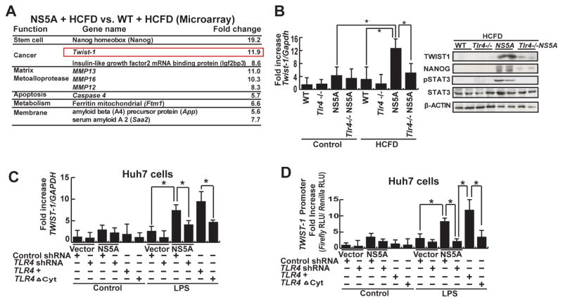
Figure 2. TLR4-mediated TWIST1 induction.
(A) Chief summary of RNA microarray analysis. Twist1, key regulator of EMT signaling was significantly higher in NS5A+HCFD compared to WT+HCFD. (B) Quantitative analysis of Twist1 from liver/liver tumor tissues of all cohorts, as listed in Figure 1A. Heightened Twist1 expression (NS5A Tg mice fed HCFD) was abrogated by TLR4 deficiency. Data normalized to GAPDH expression are listed as the fold change (*, P <0.05). (C) LPS induced TWIST1 in Huh7 cells transduced with an NS5A expression vector (*, P<0.05 compared to cells transduced with an empty vector). This was suppressed by lentiviral expression of shRNA for TLR4 and also in cells transduced with the dominant negative TLR4 vector (TLR4ΔCyt). (D) LPS induced TWIST1 promoter activity. Huh7 cells transfected with TWIST1 promoter-luciferase construct were stimulated with LPS (10 μg/ml) in culture. Other experimental procedures in this figure are the same as described earlier. TLR4 knockdown or mutation abrogated TWIST1 promoter activity, but adding TLR4 rescued it. Relative light units (RLU) values were normalized by the Renilla luciferase activity driven by SV40 promoter which were used a transfection control (*, P < 0.05).

