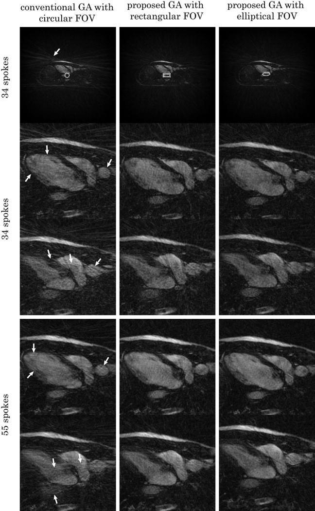
FIG. 5.

Comparison of horizontal long-axis cardiac images with different FOV shapes. Real-time acquisition with breath-hold were used for all images. Top: diastolic frame with 34 spokes reconstructed with gridding using conventional GA, proposed GA with rectangular and elliptical FOV (major-to-minor-axis ratio 1:0.4) sampling respectively. The unaliased FOV shapes are shown (white dashed contour) for each sampling scheme. The enlarged region of interest, together with another systolic frame are also shown. Bottom: two frames from the same acquisition reconstructed with 55 spokes. The conventional GA cases contain a visibly larger amount of streaking artifact due to undersampling (white arrows).
