
Fig. 2.
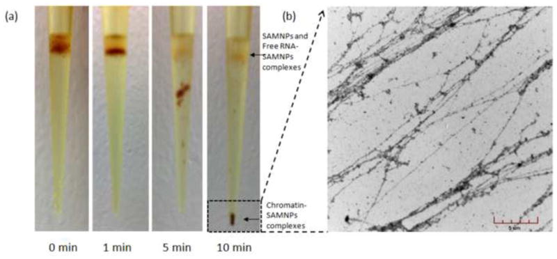
Optical images of the pipette tip during magnetophoretic chromatography of chromatin-SAMNPs in 25% (m/v) PEG solution (a). In the experiment time scale, the chromatin-SAMNPs complexes were isolated at the bottom of the tip; while the free RNA-SAMNPs and free SAMNPs were kept at the interphase of the two solutions. TEM images of chromatin released from the chromatin-SAMNPs complexes in PBS solution (b).