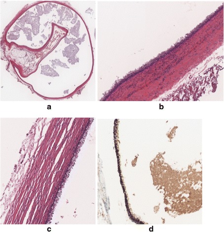
Fig. 4.

Microphotographs showing the histopathological properties of the cyst. a Overall view of the ciliated foregut cyst (haematoxylin and eosin, ×22 magnification). b Pseudostratified ciliated columnar epithelium resting on sub-epithelial connective tissue, a smooth muscle layer and an outer fibrous layer (haematoxylin and eosin, ×349 magnification). c Mucus-laden goblet cells in some areas of the cyst (haematoxylin and eosin, ×540 magnification).d CK7-positive epithelial lining (CK7 immunohistochemical stain, ×400 magnification). The epithelium is negative for CK20 and CDX2 immunohistochemical stains (not shown in the figure)
