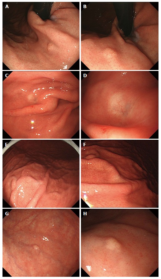
Figure 1.

Endoscopic findings: Case 1 (A, B), case 2 (C, D), case 3 (E, F) and case 4 (G, H). Conventional endoscopy on white-light imaging showed flat lesions with whitish discoloration (C-F) or yellowish submucosal tumor shapes (A, B, G, H). Dilated vessels were shown on the surface of tumors (B, D, F, H).
