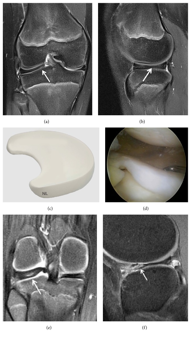
Figure 16.
Discoid lateral meniscus. (a) Coronal T2 FSE Fat Sat MRI showing meniscal enlargement. The lateral meniscal body (arrow) is enlarged and has a more slab-like configuration compared to the normal-appearing triangular medial meniscal body; (b) Sagittal T2 FSE Fat Sat image of the lateral meniscus demonstrating persistence of the bow tie appearance on the more central slices rather than converting into 2 opposing triangles; (c) three-dimensional diagram showing a discoid lateral meniscus; (d) arthroscopic views of a discoid lateral meniscus; (e) posterior cystic degeneration in a discoid lateral meniscus; (f) anterior cystic degeneration in a discoid lateral meniscus.

