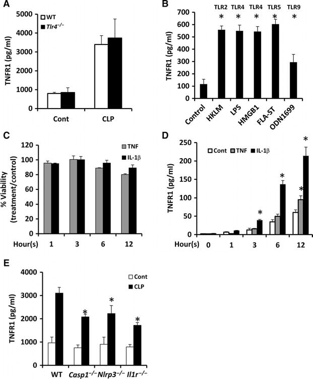
Fig. 1. Multiple TLR ligands and cytokines stimulate HC-TNFR1 shedding in vitro and in vivo.

(A) Wild-type (WT) and Tlr4−/− mice were subjected to CLP. In the control mice, laparotomy and cecal manipulation were performed without ligation and puncture. Eight hours later, plasma sTNFR1 concentrations were measured by ELISA. Data are means ± SD from 8 to 10 mice per group from two separate experiments. (B) Mouse hepatocytes were left untreated (Control) or were stimulated for 24 hours with the TLR ligands HKLM (1 × 10⁸ cells/ml), LPS (100 ng/ml), HMGB1 (5 μg/ml), FLA-ST (1 μg/ml), and ODN1669 (5 μg/ml). The concentrations of TNFR1 in the culture media were measured by ELISA. Data are means ± SD from three experiments. *P < 0.05 versus control by two-tailed unpaired t test. (C and D) Mouse hepatocytes were left untreated or were incubated with TNF (500 U/ml) or IL-1β (100 U/ml) for the indicated times. At each time point, (C) cell viability and (D) the concentration of TNFR1 in the culture media were determined. Data are means ± SD from three experiments. *P < 0.05 versus control by two-tailed unpaired t test. (E) Mice of the indicated genotypes were subjected to CLP. Eight hours later, the plasma concentrations of TNFR1 were determined by ELISA. Data are means ± SD from six mice per group of each genotype from two experiments. *P < 0.05 versus control by two-tailed unpaired t test.
