
Fig. 2. TNFR1 shedding from hepatocytes and myeloid cells during sepsis depends on Myd88.
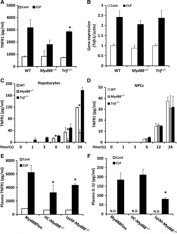
(A and B) WT, Myd88−/−, and Trif−/− mice were subjected to CLP. Eight hours later, (A) plasma sTNFR1 concentrations were determined by ELISA, and (B) Tnfr1 mRNA abundance in the liver was determined by real-time reverse transcription polymerase chain reaction (RT-PCR) analysis. Data are means ± SD from 8 to 10 mice per group from two experiments. *P < 0.05 versus control by two-tailed unpaired t test. (C and D) Hepatocytes (C) and NPCs (D) from the indicated mice were left untreated (zero-hour time point) or were treated with LPS for the indicated times. At each time point, the concentration of TNFR1 in the culture media was determined by ELISA. Data are means ± SD from three experiments. *P < 0.05 by one-way analysis of variance (ANOVA). (E and F) Myd88flox, HC-Myd88−/−, and LysM-Myd88−/− mice were subjected to CLP. Eight hours later, plasma concentrations of (E) TNFR1 and (F) IL-1β were determined by ELISA. Data are means ± SD from 8 to 10 mice per group from two experiments. *P < 0.05 versus Myd88flox by two-tailed unpaired t test. N.D., not detectable.