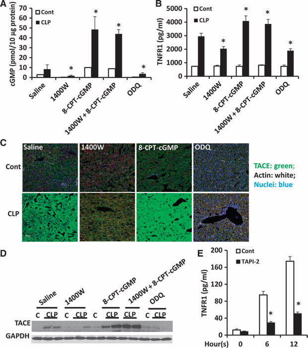
Fig. 4. iNOS-stimulated HC-TNFR1 shedding during polymicrobial sepsis is mediated by cGMP and TACE.

(A to D) WT mice were subjected to CLP and then were immediately treated subcutaneously with saline, 1400W (5 mg/kg), 8-CPT-cGMP (5 mg/kg), both 1400W and 8-CPT-cGMP, or ODQ (20 mg/kg). Eight hours after CLP, the concentrations of (A) cGMP in liver homogenates and (B) TNFR1 in plasma were determined. Data are means ± SD from six to eight mice per group from two experiments. *P < 0.05 by one-way ANOVA. (C) Some of the same mice were analyzed by immunofluorescence microscopy to detect active TACE in the liver. TACE is shown in green, nuclei are in blue, and actin is shown in white. Scale bar, 50 μm. (D) Liver homogenates from the same mice were analyzed by Western blotting to detect TACE. GAPDH was used as a loading control. Western blots are representative of two independent experiments. (E) Hepatocytes isolated from WT mice were left untreated (zero-hour time point) or were treated in vitro with LPS (100 ng/ml) in the absence or presence of 400 nM TAPI-2 for the indicated times. The concentration of TNFR1 in the culture media was determined by ELISA. Data are means ± SD from three independent experiments. *P < 0.05 by one-way ANOVA.
