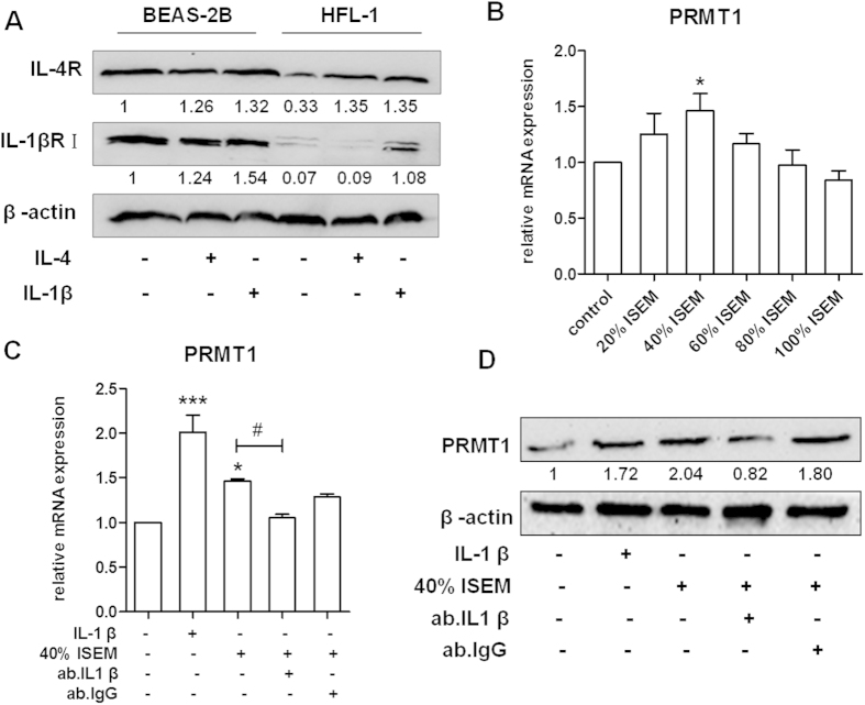
Figure 3. Expression of IL-4 receptor and IL-1β receptor I in BEAS-2B and HFL-1 cells and expression of PRMT1 after stimulation with ISEM in HFL-1 cells.
The expression of IL-4 receptor and IL-1β receptor I were detected by Western blotting with or without IL-4 and IL-1β stimulation in BEAS-2B and HFL-1cells (A). The supernatant from IL-4-stimulated BEAS-2B cells (ISEM) was accumulated after 48 hours stimulation and 5% FBS was added to the culture supernatant to replenish the consumption by epithelial cells. Then ISEM was used to stimulate HFL-1 cells. The expression of PRMT1 in HFL-1 cells was determined by RT-qPCR after 24h stimulation by different concentrations of ISEM (20%, 40%, 60%, 80%, 100%) (B). IL-1β, 40% ISEM, and IL-1β antibody were used to stimulate HFL-1 cells and the expression of PRMT1 was detected by RT-qPCR and Western blotting (C,D). Western blotting was shown as representative image and density under the band was measured (ImageJ software) and normalized to β-actin. The results were expressed as mean ± S.E.M of triplicates from three independent experiments and analyzed by One-way ANOVA test. *,# and ** represent P < 0.05 and P < 0.01 between indicated groups and control group.

