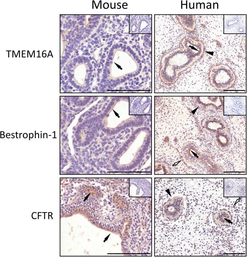
Figure 1. Expression of chloride channels in the developing mouse lungs and human fetal lungs.
Paraffin-embedded, 5 μm-thick sections from E12.5 mouse (left panel) and week 9–11 human fetal lungs (right panel) were dewaxed and used for immunohistochemistry. Expression of the Ca2+-activated chloride channels TMEM16A, bestrophin-1 and CFTR were visualised using DAB (brown straining) in the lung epithelium. Sections were counterstained with Harris’ hematoxylin (blue staining). Negative controls were carried out in serial sections form the same lungs by substituting the primary antibody with an isotype control (inset). Block arrows show apical expression in the epithelium, arrowheads show basolateral expression and open arrows show expression in the mesenchyme. Scale bar = 100 μm.

