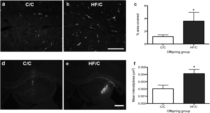
Figure 6.
(a–c) Photomicrographs of fibronectin staining in the hippocampus of 11-month old C/C (a) and HF/C (b) Tg2576 mice. Fibronectin covered a significantly greater area in HF/C mice compared to C/C animals (c). (d,e) Photomicrographs of exogenous HilyteFluor-488 human Aβ40 injected into the hippocampus of 5-month old wildtype C/C (d) and HF/C mice (e). (f) Significantly more Aβ was retained at the injection site in the hippocampi of HF/C mice, as indicated by the increased mean fluorescence intensity measured at the site of injection. Scale bars: a and b = 500 μm, d and e = 400 μm. *p < 0.05.

