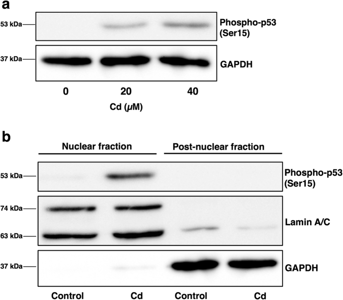
Figure 5. Cd increases phosphorylated p53 protein levels in HK-2 cells.
(a) Phosphorylated p53 protein levels in Cd-treated HK-2 cells. Whole cell lysates of HK-2 cells treated with 20 or 40 μM Cd (CdCl2) for 6 h were used for western blot analysis and probed with phospho-p53 antibody. Anti-GAPDH antibody was used as a loading control. Upper panel, phosphor-p53; lower panel, GAPDH. The two blots were run under the same experimental conditions. Uncropped images are provided in Supplementary Fig. 1f. (b) Localization of phosphorylated p53 protein in Cd-treated HK-2 cells. HK-2 cells were treated with 40 μM Cd for 3 h and separated into nuclear and post-nuclear fractions. Western blotting for the detection of phospho-p53 was performed (upper panel). Lamin A/C, nuclear marker (middle panel); GAPDH, cytosolic marker (lower panel). The three blots were run under the same experimental conditions. Uncropped images are provided in Supplementary Fig. 1g.

