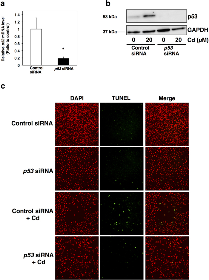
Figure 6. Knockdown of p53 decreases Cd-induced apoptosis in HK-2 cells.
(a) Knockdown efficiency of p53 in HK-2 cells following p53 siRNA treatment. p53 siRNA was added to HK-2 cells and incubated for 24 h. mRNA levels were examined using real-time RT-PCR. mRNA levels were normalized to GAPDH. Values are the mean ± S.D. (n = 3). *Significantly different from the control group, P < 0.05. (b) Protein levels of p53 in HK-2 cells treated with p53 siRNA and Cd (CdCl2). HK-2 cells were treated with or without p53 siRNA for 24 h, and then treated with or without 20 μM Cd for 18 h. Whole cell lysates of HK-2 cells were used for western blot analysis. Anti-GAPDH antibody was used as a loading control. Upper panel, p53; lower panel, GAPDH. The two blots were run under the same experimental conditions. Uncropped images are provided in Supplementary Fig. 1h. (c) Representative images of TUNEL-positive apoptotic cell (green signals). HK-2 cells were treated with or without p53 siRNA for 24 h, and then treated with or without 20 μM Cd for 18 h. Nuclei were counterstained with DAPI, and the blue signal was converted to red (DAPI). Fluorescein signals from TUNEL-positive cells (TUNEL) and red signals of nuclei were merged (Merge). Original magnification: × 100.

