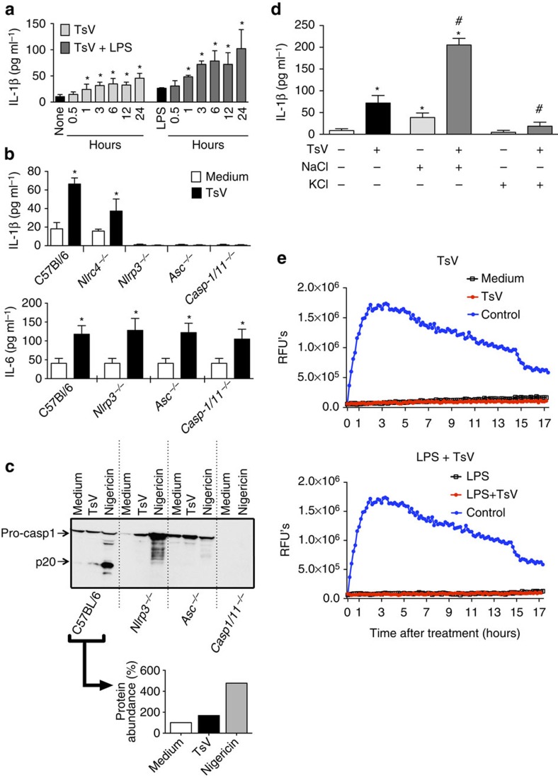
Figure 1. T. serrulatus venom (TsV) induces the activation of the NLRP3 inflammasome in macrophages.
BMDMs from C57Bl/6 mice were pre-treated or not (a) with LPS (1,000 ng ml−1) for 4 h and then stimulated with TsV (50 μg ml−1) for 0.5, 1, 3, 6, 12 and 24 h to induce IL-1β release, which was measured by ELISA. (n=4) (b) IL-1β and IL-6 production measured by ELISA in the culture supernatant of BMDMs from C57Bl/6 WT, Nlrc4−/−, Nlrp3−/−, Asc−/− and Casp1/11−/− mice; the cells were incubated only with TsV (50 μg ml−1) for 24 h. (n=4) (c) C57Bl/6 WT, Nlrp3−/−, Asc−/− and Casp1/11−/− BMDMs were pre-treated for 4 h with LPS (1,000 ng ml−1) and later stimulated with TsV (50 μg ml−1) for 24 h or nigericin as a positive control (20 μM) for 1 h. Immunoblotting for caspase 1 (p20) in the culture supernatant was performed and the percentage of p20 abundance was calculated by densitometric analysis and is shown next to the panels. The data are representative of two independent experiments. (d) BMDMs from C57BL/6 mice were stimulated with TsV (50 μg ml−1) plus NaCl (50 mM) or KCl (50 mM) for 24 h and IL-1β was detected in the cell supernatant by ELISA. All data are representative of three independent experiments (n=3). (e) BMDMs from C57Bl/6 mice were pre-treated or not with LPS (1 μg ml−1) for 4 h and then stimulated with TsV (50 μg ml−1) or Triton-X (9%), which was used as the positive control for pore formation. Fluorometric plots show propidium iodide uptake (RFUs) over time to demonstrate the kinetics of pore formation in cell membrane. The data are representative of two independent experiments, performed in triplicate. Error bars (a,b,d), s.d. *P<0.05 (one-way ANOVA with Bonferroni's post-test compared with non-TsV-stimulated), #P<0.05 (one-way ANOVA with Bonferroni's post-test compared with TsV-stimulated). Images have been cropped for presentation. Full size images are presented in Supplementary Fig. 6. RFU, relative fluorescence units.

