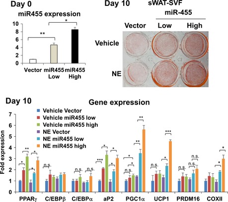
Figure EV1. miR‐455 induced brown adipogenesis of primary sWAT‐SVFs.

SVF was isolated from subcutaneous white adipose tissue (sWAT) of C57BL/6 mice, plated in a cell culture dish, and transduced with different dosages of miR‐455 and control (vector) lentiviruses to achieve different levels (low or high) of miR‐455 overexpression. At confluence, the cells were induced to differentiate by standard differentiation protocol (see Materials and Methods) with supplement of 1 mM rosiglitazone. On day 10, cells were treated with 100 mM norepinephrine (NE) for 4 h and stained with Oil Red O, and RNA was isolated for gene expression analysis by qRT–PCR. Data were analyzed with Student's t‐test and are presented as mean ± SEM of a representative from three independent experiments each performed in quadruplicates (*P < 0.05, **P < 0.01, and ***P < 0.001; n.s., non‐significant).
