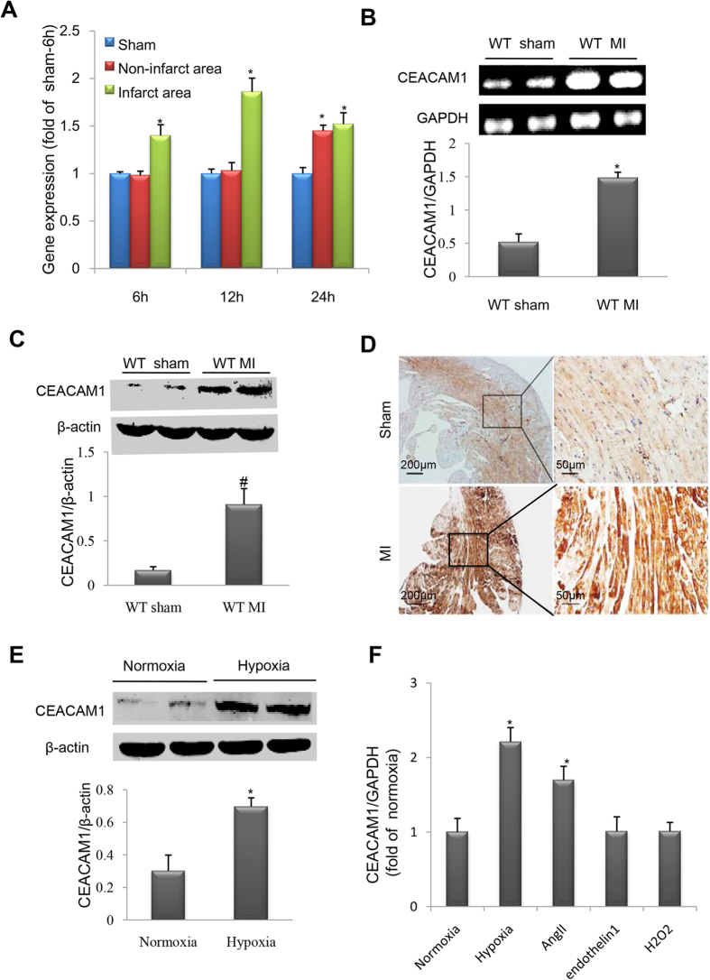
Figure 2. Myocardial CEACAM1 was upregulated in response to myocardial infarction (MI) or hypoxia.
(A), Time course of CEACAM1 mRNA level in sham, non-infarct area and infarct area. *P < 0.05 vs. sham 6h. Expression of CEACAM1 mRNA (B) and protein (C) in the left ventricle of wild-type (WT) mice with MI for 3 days (n = 4 in each group). *P < 0.01 vs. sham. (D), Immunohistochemical staining shows elevated myocardial CEACAM1 expression in mice with MI than in mice with sham operation (non-infarct area). (E), Immunoblotting of CEACAM1 expression in neonatal rat cardiomyocytes exposed to hypoxia for 24 hours (n = 5 in each group). *P < 0.01 vs. normoxia. (F), The CEACAM1 mRNA level in neonatal rat cardiomyocytes exposed to hypoxia (for 3h), angiotensin II (AngII, 1 umol/L), endothelin 1(0.1 umol/L) and H2O2 (0.15 umol/L) for 24 hours. *P < 0.05 vs. normoxia.

