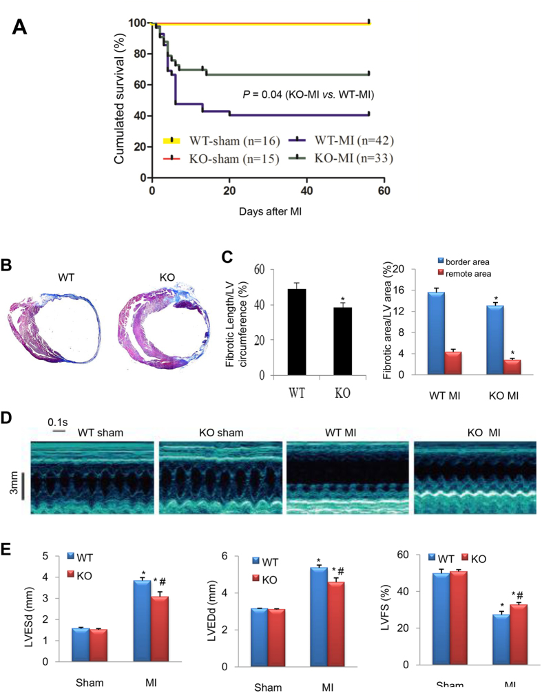
Figure 3. Ablation of CEACAM1 improved the survival rate and post-MI cardiac remodeling.
(A), Wild-type (WT) and CEACAM1 knockout (KO) mice were subjected to sham operation or MI. Then survival was monitored for 8 weeks. (B) Masson trichromatic staining of WT and KO mouse hearts at 8 weeks after MI (blue indicates collagen). (C) Quantification of the length of fibrosis in the infarct area (relative to the total LV circumference) and the fibrotic area in the border zone and the remote area. *P < 0.05 vs. the corresponding WT group, n = 6 in each group. (D) Representative echocardiographic images from the four groups. (E) Quantification of left ventricular end-systolic and end-diastolic diameter (LVESd, LVEDd), and left ventricular fractional shortening (LVFS). *P < 0.01 vs. corresponding sham group, #P < 0.05 vs. WT MI. (n = 4 in sham groups; n = 9 in MI groups).

