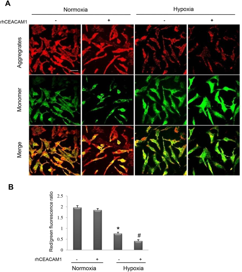
Figure 7. Influence of recombinant human CEACAM1 (rhCEACAM1) on the mitochondrial membrane potential (ΔΨm) in neonatal rat cardiomyocytes exposed to hypoxia for 24 h.
JC-1 fluorescent dye in the mitochondrialmatrix produces red fluorescence. If the mitochondrial membrane potential (ΔΨm) declines, JC-1 becomes a monomer in the matrix and produces green fluorescence. (A), Representative pictures of JC-1 fluorescence (indicating mitochondrial depolarization) detected by laser confocal microscopy. Scale bar, 20 μm. (B), Quantitative analysis of fluorescence intensity in the mitochondria of cardiomyocytes shown in A. RhCEACAM1 decreased the ΔΨm of hypoxic cardiomyocytes (n = 4 in each group). *P < 0.01 vs. normoxia and #P < 0.05 vs. hypoxia.

