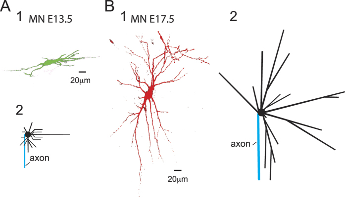
Figure 1. Morphology of E13.5 and E17.5 MNs and their canonical counterparts that were used in the simulations.
(A1) Photograph of a neurobiotin-filled E13.5 MN as revealed with fluorescent dye. (A2) Morphology of the canonical E13.5 MN that was used in the simulation. (B1-2) The same disposition as in (A1-2) but for E17.5 MNs.

