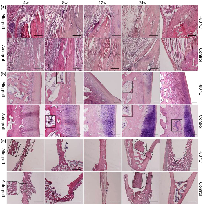
Figure 1. Photomicrographs of the junction of the quadriceps tendon and the soft tissues, patella and tibial insertion site.
H&E staining of the junction of the quadriceps tendon and the soft tissues (a) (scale bar, 1 mm), the patella (b) (scale bar, 200 μm) and the tibial insertion site (c) (scale bar, 1 mm) in the allograft and autograft groups at 4, 8, 12 and 24 weeks postoperatively and in the −80 °C and control groups. Inserts show higher magnification of vessels in (b) and higher magnification of tibial insertion sites in (c).

