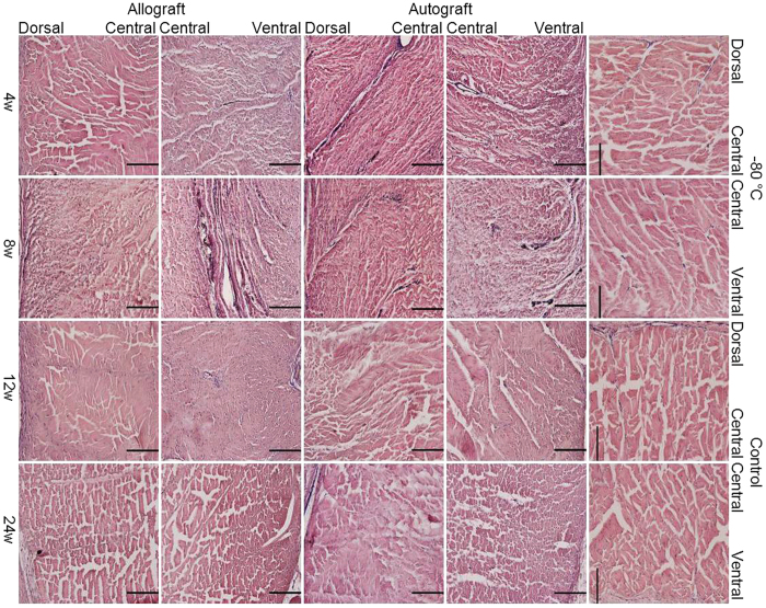
Figure 4. Robust but delayed revascularization in allograft patellar tendon.
H&E staining of the dorsal, central and ventral portions of the middle part of the patellar tendon (transverse section) after India-ink perfusion in the allograft and autograft groups at 4, 8, 12 and 24 weeks postoperatively and in the −80 °C and control groups. The scale bar represents 200 μm.

