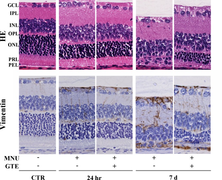
Fig. 2.
Retinal morphology and vimentin immunoreactivity in normal animals and those treated with N-methyl-N-nitrosourea (MNU) (24 hr and 7 days), with or without green tea extract (GTE). Retinal regions depicted are as follows: GCL, ganglion cell layer; IPL, inner plexiform layer; INL, inner nuclear layer; OPL, outer plexiform layer; ONL, outer nuclear layer; PRL, photoreceptor layer; PEL, pigment epithelial layer.

