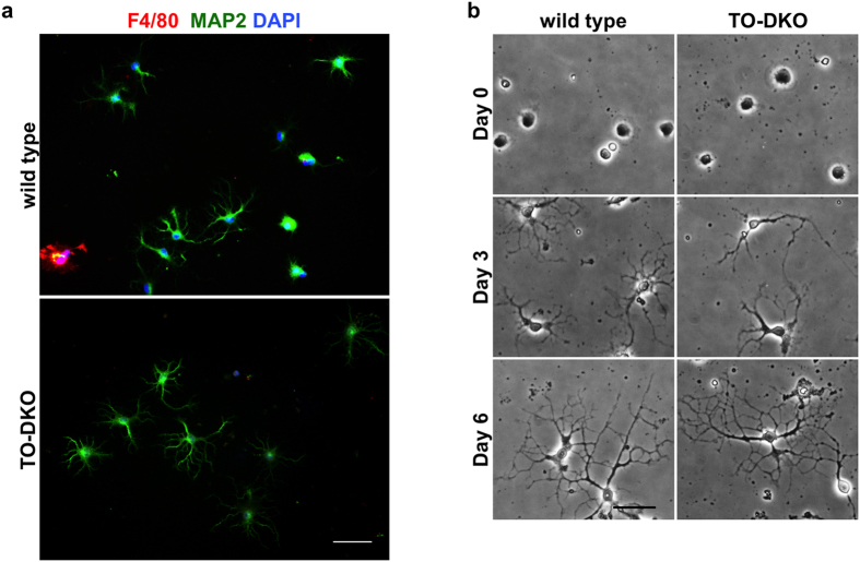
Figure 2. Cortical neurons isolated from adult wild-type and Mth1/Ogg1-DKO brains regenerate neurites.
(a) Purity of cortical neuron culture at day 3 in vitro. Neuronal (MAP2, green) and microglial (F4/80, red) markers were detected by immunofluorescence microscopy. Nuclei were counter stained by DAPI (blue). About 95% of the cultured cells were MAP2-positive neurons. Merged images are shown. Scale bar = 50 μm. (b) Phase contrast images of cortical neurons isolated from adult wild-type (left panels) and Mth1/Ogg1-DKO (TO-DKO) (right panels) mice cultured for 0, 3 and 6 days, showing time-dependent neurite regeneration. Scale bar = 20 μm.

