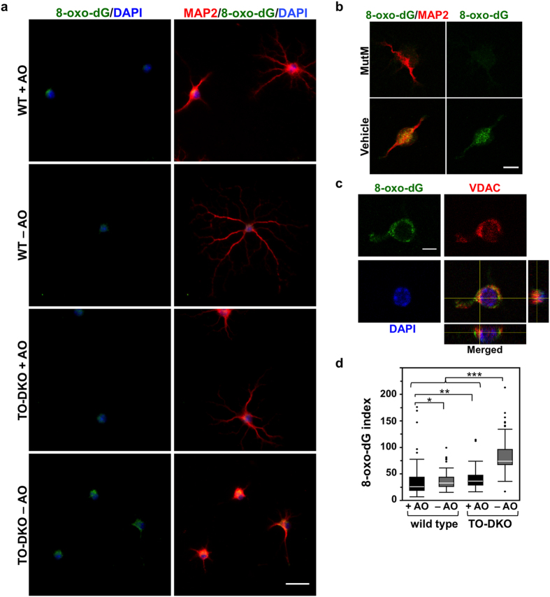
Figure 6. MTH1/OGG1 deficiency significantly increased accumulation of 8-oxoguanine in the mitochondrial DNA of cortical neurons in the absence of antioxidants.
(a) 8-Oxo-deoxyguanosine (8-oxo-dG) detected in MAP2-positive neurons by immunofluorescence microscopy. Fixed neurons pre-treated with RNase were subjected to a mild denaturation with 25 mM NaOH before reacting with antibodies. Adult cortical neurons isolated from Mth1/Ogg1-DKO (TO-DKO) and wild-type (WT) mice were cultured for 2 days in the absence (−AO) or presence (+AO) of antioxidants. Green: 8-oxo-dG; red: MAP2: blue: DAPI. Scale bar = 20 μm. Cytoplasmic 8-oxo-dG immunoreactivity was increased in TO-DKO neurons maintained in the absence of antioxidant. (b) 8-Oxo-dG immunoreactivity in TO-DKO neurons in the absence of antioxidants was completely abolished by pre-treatment with MutM 8-oxoG DNA glycosylase. Scale bar = 10 μm. (c) Mitochondrial localization of 8-oxo-dG in a TO-DKO neuron. Immunofluorescence signals for mitochondrial voltage-dependent anion channel (VDAC, red) were co-localized with the cytoplasmic 8-oxo-dG immunofluorescence (green). Orthogonal views obtained by laser scanning confocal microscopy are shown. Blue: DAPI. Scale bar = 10 μm. (d) Quantitative evaluation of mitochondrial 8-oxo-dG in adult cortical neurons with (+AO) or without (−AO) antioxidants. More than 203 cells were examined for each group. 8-Oxo-dG indexes were calculated and are presented as whisker-box plots. Outliers are shown as dots. Wilcoxon/Kruskal–Wallis tests, chi square test p < 0.0001; post hoc comparison with Wilcoxon method, *p < 0.05, **p < 0.005, ***p < 0.0001.

