
Figure 2. Displacements in the CYP101 structure upon removal of substrate from the active site binding pocket (haem, in red).
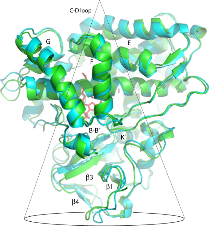
Substrate-free CYP101 (2LQD) is shown in cyan, while the camphor-bound form is in green (2L8M)9,10. Relevant secondary structure elements are labeled. Many of the largest displacements occur within the conical volume shown traced in black. See also Video 2.