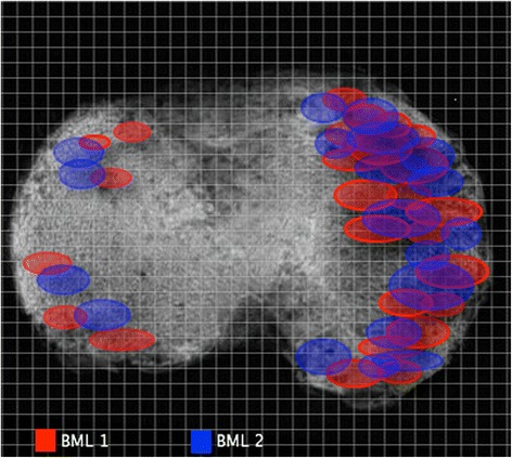
Fig. 2.

The approximate external contour of each bone marrow lesion (BML) area was marked. The precise map location was placed by identifying the number of sagittal and coronal slices with measurement of distance from the external tibial contour. After marking the position of the BML for all specimens, a distribution map of both types of BML was found. BML 1 bone marrow lesion detected using the fast spin-echo proton density-weighted sequence only, with absent signal on T1-weighted sequence in the same area, BML 2 bone marrow lesions detected by both fast spin-echo proton density-weighted and T1 sequences
