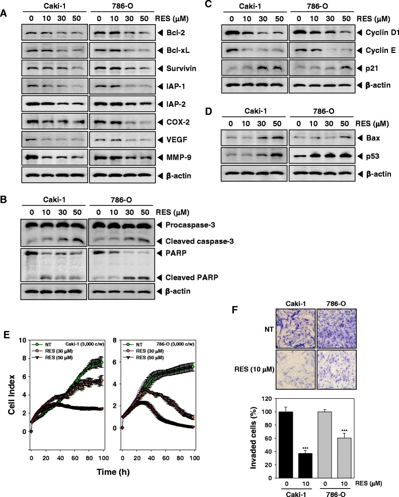
Fig. 4.

RES suppresses expression of various proteins involved in anti-apoptosis, proliferation, and angiogenesis. a-d After Caki-1 and 786-O cells (1 × 10⁶ cells/well) were incubated with the indicated various concentrations of RES for 24 h. Whole-cell extracts were prepared, and 20 μg of the whole-cell lysate was resolved by SDS-PAGE, electrotransferred to nitrocellulose membrane, and then probed with antibodies against bcl-2, bcl-xL, survivin, IAP1/2, COX-2, VEGF, MMP-9, caspase-3, PARP, cyclin D1, cyclin E, p21, Bax, and p53 as described in “Materials and methods.” The same blots were stripped and reprobed with β-actin antibody to verify equal protein loading. The results shown here are representative of three independent experiments. e Cell proliferation assay was performed using the Roche xCELLigence Real-Time Cell Analyzer (RTCA) DP instrument (Roche Diagnostics GmbH, Germany) as described in “Material and methods”. After Caki-1 and 786-O cells (5 × 10³ cells/well) were seeded onto 16-well E-plates and continuously monitored using impedance technology. f Caki-1 and 786-O cells were added to the upper side of the invasion chamber, the cells on the upper surface of the filter were removed after 24 h of incubation in the presence or absence of 10 μM of RES. The cells on the lower surface were fixed, stained and monitored by photographing, then counted. Randomly chosen fields were photographed under a light microscope at 100× magnification and invaded cells were counted
