Fig. 1.

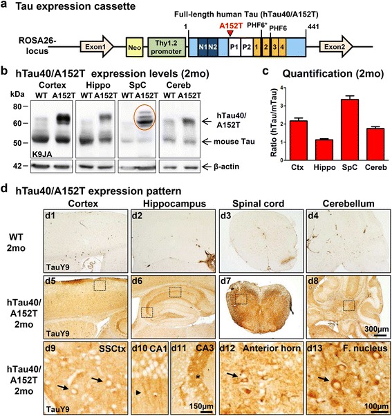
Generation of human hTau40AT mice with the Tau mutation A152T. a Illustration of the mutated hTau40AT -gene located in the ROSA26-locus. Scheme represents human full-length Tau (hTau40) with highlighted A152T mutation (red), proline-rich domain (white, P1, P2), N-terminal inserts (dark blue, N1, N2) and aggregation-prone hexapeptide motifs (PHF6*, PHF6) in the repeat domain (orange, 1–4). hTau40AT -expression is controlled by the murine Thy1.2 promoter. b Representative expression of hTau40AT (Mr ~67 kDa) and endogenous mouse Tau (Mr ~ 45-55 kDa) in cortex, hippocampus, spinal cord and cerebellum of 2 months old hTau40AT mice and age matched controls (WT) detected by the pan-Tau antibody K9JA. Note the slightly up-shifted human Tau in the spinal cord sample caused by hyperphosphorylation (red circle). ß-actin serves as loading control. c Quantification of (b). Ratio of hTau40AT to endogenous mTau indicates the hTau40AT expression level in different brain regions, e.g. cortex (~2x), hippocampus (~1x), spinal cord (~3x) and cerebellum (~1.5x). Each bar shows mean ± SEM of n = 17 animals. d Uniform distribution of hTau40AT visualized by the human Tau specific antibody TauY9 in brain (cortex, hippocampus and cerebellum) and spinal cord of 2 months old hTau40AT mice (d5-d8). Boxed areas with higher magnification (d9-d13) indicate mislocalization of hTau40AT into cell bodies (arrows) and dendrites (arrowhead) and a strong immunoreactivity of mossy fibers in the CA3 hippocampal area (asterisk) in comparison to WT mice (d1-d4). WT: wild-type; A152T: hTau40AT transgenic mouse strain; Ctx: cortex; Hippo: hippocampus; SpC: spinal cord; Cereb: cerebellum; SSCtx: somatosensory cortex; CA: cornu ammonis; F. nucleus: Fastigial nucleus; mo: months; Scale bars: 100 μm (d9, d10, d12, d13), 150 μm (d11), 300 μm (d1-d8)
