Fig. 2.

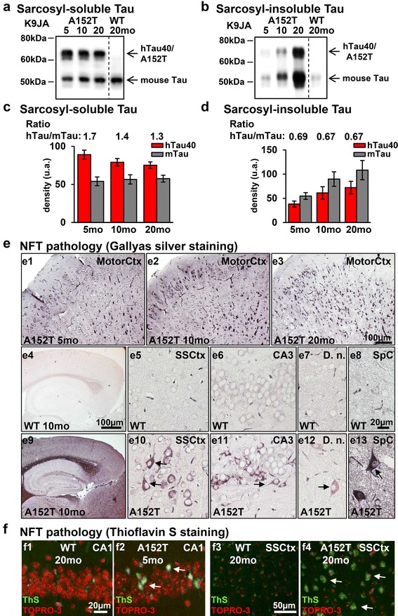
Progressive co-aggregation of endogenous mouse Tau and exogenous human hTau40AT in mice. a-b Western blot analysis using the pan-Tau antibody K9JA shows sarcosyl-soluble and sarcosyl-insoluble Tau species of hTau40AT (upper band; Mr ~67 kDa) and mouse Tau (lower band; Mr ~45-55 kDa). Note (a) the slight reduction of soluble hTau40AT and (b) a progressive co-aggregation of insoluble mouse and human Tau in aging hTau40AT mice. c Quantification of (a). Densitometric measurements of hTau40AT (red bars) and mouse Tau (grey bars) indicate a reduction of soluble hTau40AT with age (5-20mo) (note the declining ratio in aging transgenic mice). Each bar shows mean ± SEM of n = 4 animals. d Quantification of (b). Densitometric measurements of hTau40AT (red bars) and mouse Tau (grey bars) indicate an increase of insoluble human hTau40AT and mouse Tau with increasing age (5-20mo). Each bar shows mean ± SEM of n = 4 animals, error bars represent SEM. e Gallyas silver staining of different brain areas. (e1-e3) Progressive Tau aggregation in motor cortex of hTau40AT mice with increasing age. Note NFTs with flame-shaped structure (arrows) in cortical (e10), hippocampal (e11), cerebellar (e12) and spinal (e13) neurons of 10 months old hTau40AT mice, compared to silver-negative control (e4-e8). f Thioflavin S staining (green) and counterstaining with TOPRO-3 (red) of hippocampal CA1 pyramidal neurons (f2, arrows) and somatosensory cortical neurons (f4, arrows) confirms the presence of NFTs in hTau40AT mice at 5 and 20 months of age and the absence of Tau-aggregates in WT mice (f1, f3). WT: wildtype; A152T: hTau40AT transgenic mouse strain; SSCtx: somatosensory cortex; SpC: spinal cord; D.n.: Dentate nucleus; CA: cornu ammonis; mo: months; Scale bars: 20 μm (e5-e8, e10-e13, f1-f2); 50 μm (f3-f4); 100 μm (e1-e4, e9)
