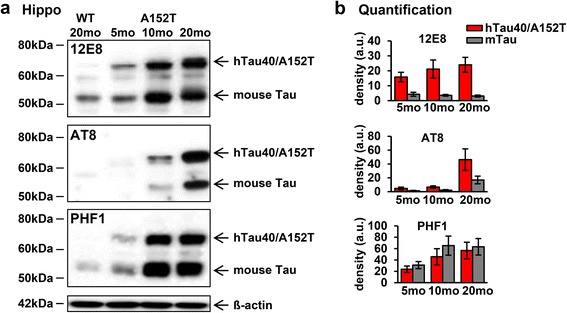
Fig. 4.

Progressive hyperphosphorylation of Tau in aging hTau40AT mice (5-20mo). a Western blot analysis using phospho-Tau antibodies (12E8, AT8, PHF1) on hippocampal extracts of 5–20 months old hTau40AT compared to 20 months old WT mice. b Quantification of (a). Densitometric analysis of hTau40AT (red bars) and mouse Tau (grey bars) indicate an increase of hyperphosphorylated human hTau40AT and mouse Tau in aging hTau40AT mice (5-20mo). Each bar shows mean ± SEM of n = 4 animals. ß-actin serves as loading control. WT: wild type; A152T, hTau40AT transgenic mouse strain; mo: months, a.u.: arbitrary units
