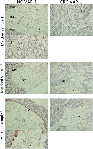
Fig. 3.

Paraffin-embedded sections from 3 matched normal colon (NC) and CRC samples demonstrating positive VAP-1 staining in the lamina propria and submucosa of the colon with only weak staining in CRC stroma. Magnification x200. VAP-1 staining is brown-red, haematoxylin staining is blue. MM = muscularis mucosae; SM = submucosal; LP = lamina propria; St = tumour stroma; Epi = tumour epithelium
