Fig. 4.

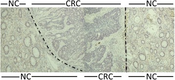
VAP-1 expression in flanking normal colon (NC) tissue abruptly disappears at the border with CRC tissue. Dot-dash line marks the border between NC and CRC. Magnification x200. VAP-1 staining is brown-red, haematoxylin staining is blue

VAP-1 expression in flanking normal colon (NC) tissue abruptly disappears at the border with CRC tissue. Dot-dash line marks the border between NC and CRC. Magnification x200. VAP-1 staining is brown-red, haematoxylin staining is blue