Fig. 5.

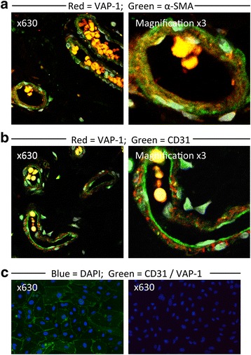
Dual-colour immunofluorescent staining of sections of normal colon tissue and cultured colonic endothelial cells. Magnification x630. a VAP-1 staining is present in SMA+ cells of vessels in sections of colonic mucosa. b VAP-1 staining does not co-localise with CD31+ endothelial staining of vessels in sections of colonic mucosa. Right-most images are digitally magnified x3. Yellow staining represents autofluorescence from erythrocytes. c Positive CD31 staining (left) and absent VAP-1 staining (right) of cultured colonic endothelial cells
