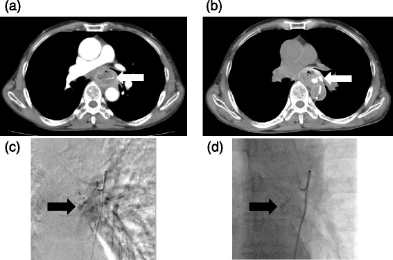
Fig. 1.

Arterial esophageal bleeding in case 2. a Enhanced CT shows that contrast material was in the esophagus in the arterial phase, indicating extravasation of contrast material (arrow). b Enhanced CT with angiography of the esophageal artery shows contrast material extravasation in the esophagus (arrow). c An image of digital subtraction angiography of the esophageal artery demonstrates punctate contrast material collections in the esophagus (arrow). d After transcatheter arterial embolization with a mixture of N-butyl cyanoacrylate (NBCA) and Lipiodol (arrow), active bleeding improved
