
Figure 6.
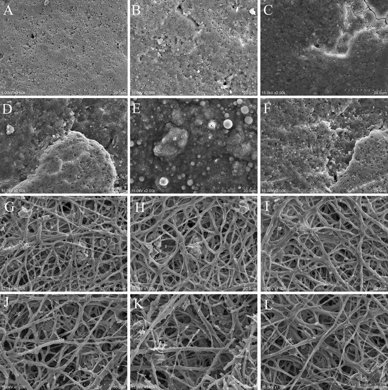
Scanning electron microscopy of the inner and outer surface of the eggshells of laying hens at different times post inoculation with AIV. Representative scanning electron microscopy photograph of the outer (A–F) and inner surface (G–L) of the eggshells from the control hens (A, G) and in hens at 1~7 days post inoculation with AIV (B–F, H–L).