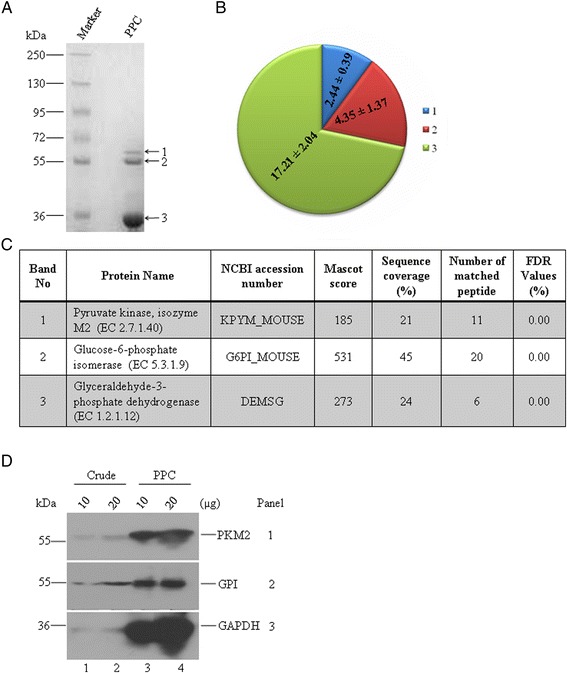
Fig. 1.

GAPDH is associated with PKM2 and GPI. a A representative of coomassie blue stained 7.5–15 % Tris-glycine SDS-PAGE gel of 10 μg purified protein complex (PPC) from EAC cells. Three major bands with different molecular weight approximately 58, 55, 33 kDa are labeled with 1, 2, and 3, respectively. b Mole of a subunit in the complex was calculated using a formula = [{(intensity of the band/total intensity of three bands) x loading amount}/molecular mass]. Molar ratio is shown in pie chart. c Three bands were cut out from the gel for trypsin digestion followed by MALDI-TOF/TOF analyses. Score of identified proteins from each band is tabulated. Note that bands 1, 2 and 3 contain mainly PKM2, GPI and GAPDH respectively. Data from one representative experiment is shown here. Experiment was repeated six times. d Immunoblots of two different amounts of crude extract and purified protein complex (PPC) from EAC cells with antibodies specific for PKM2 (panel 1), GPI (panel 2) and GAPDH (panel 3). Note that both PKM2 and GPI were co-purified with GAPDH
